Macular Hole
Please note, this page is printable by selecting the normal print options on your computer.
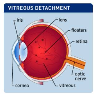
The eye is like a camera. It has a lens system at the front and a light sensitive layer which lines the back. The lens of the eye which sits just behind the coloured part (the iris), focuses the light coming through the clear window of the eye (the cornea) onto the light sensitive layer (the retina). The retina sends information to the brain through the optic nerve.
At the back of the eye in the centre of the retina the light sensitive cells are packed close together – you use this retina to see sharp detail like car number plates, people’s faces and print. It is called the fovea and here the retina is very thin. The cavity of the eyeball is filled with a jelly called vitreous. When we are young it is attached firmly all over the retina like jelly in a bowl. As we age, the vitreous breaks away from the retina leaving a layer still attached, like turning the bowl upside down and shaking the jelly – most will fall out but a layer will stay stuck to the bowl.
Fig. 1 Vitreous Separation
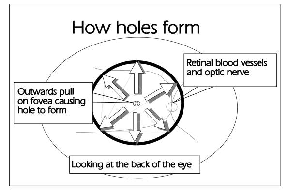
For reasons we don’t understand this vitreous remnant may shrink and because it is stuck firmly to the retina it pulls on it. Sometimes it causes a wrinkling of the retina, sometimes it pulls so hard that the thin fovea is torn and a round hole opens up. This is called a macular hole.
Fig. 2: How holes form
The first sign of the pulling is distortion of sight – straight lines appear kinked. Then the vision may get worse until even the large letters on the sight test chart are blurred and wherever you try
to look you see a grey blob.
The time it takes for the hole to form and vision to deteriorate varies considerably from person to person and from weeks to years. There is no way of predicting who will be severely affected. In some cases the vitreous remnant frees itself, the hole resolves and the vision improves. A vitrectomy operation can be done to treat this problem. The detail of the procedure depends on what stage the hole has reached. In the early stages a simple operation is done to remove the main bulk of the vitreous and to tease off the gel remnant from the retina. If the hole is at a more advanced stage, a drop of blood clotting cells (platelets) is used to glue the hole edges together and the eye is filled with a gas bubble at the end of the operation.
If gas is used you will be told to position your head face down for most of the time until the bubble disappears – usually 10 days. This is most important to make the operation work and to stop a cataract forming in the lens. (The word cataract means clouding of the lens of the eye). If you cannot manage to posture after the operation it is not worth having it done. The operation is done through three small openings in the white of the eye under general anaesthetic. A general anaesthetic is not safe for some people because of other medical problems and carries a very small risk for all.
The operation takes between one and two hours. Afterwards the eye may be a little sore or irritable for a few days, but not very painful. There are two important don’ts:
• Don’t bang or rub your eye; you may do serious damage
• Don’t do vigorous activity for two weeks.
You must:
• Keep the eyelids clean with a lint or cotton wool ball moistened with boiled, cool water
• Put your eye drops in four times daily for three weeks – get more if you run out
• Put on your glasses carefully to avoid poking the eye
• Wear glasses or sunglasses to protect the eye
• Report to the hospital or ring the emergency number straight away if within the first five days of surgery the eye becomes increasingly painful and the sight gets increasingly dim or if you do
bang it and it becomes watery
• If you were using eye drops before, check if these are to continue.
You may:
• Bath or shower, but avoid getting soapy water into the eye straight away.